Neurological Care for Kids
The neurosciences team at Children's takes care of more pediatric neurological patients than any other hospital in the country—confirming our expertise in a wide variety of neurological conditions.
View our rankings
What is neuroscience?
Neuroscience specializes in medical and surgical treatment for conditions and injuries of the nervous system and brain. Ranked among the top pediatric neurosciences programs in the country by U.S. News & World Report, our specialists at Children's Healthcare of Atlanta are uniquely qualified to help evaluate and treat pediatric neurological conditions.
Our team of neuroscience specialists has more inpatient neurology and neurosurgery visits within its program than any other pediatric hospital in the country. More visits mean more experience with a very wide variety of conditions, from the more common to the most complex. The size of our healthcare system allows us to work closely with other specialists to deliver complete, effective care for your child.
What pediatric brain conditions do we treat?
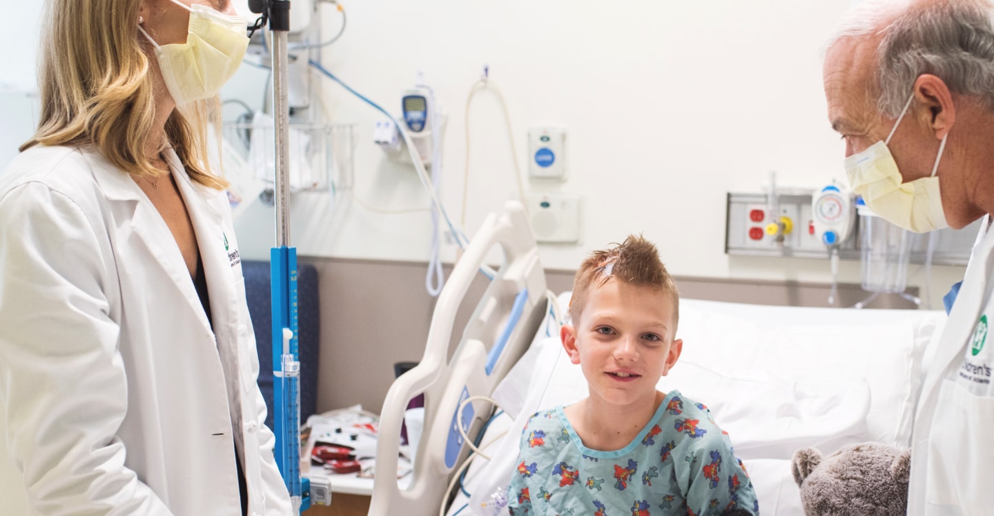
Each child deserves care tailored to his specific needs by a team that is 100% dedicated to pediatric patients.
As one of the leading pediatric neuroscience programs in the country, we specialize in medical and surgical treatment for conditions and injuries of the brain and nervous system. These include:
- Arteriovenous malformation (AVM)
- Benign subdural hygromas
- Brain abscesses and tumors
- Chiari malformation
- Concussions
- Congenital brain abnormalities
- Craniosynostosis and associated cranial abnormalities
- Epilepsy/seizure disorders
- Head injury
- Headaches
- Hydrocephalus
- Infantile spasms
- Intraventricular hemorrhage
- Large head (macrocephaly)
- Moyamoya disease
- Skull fractures
- Sleep disorders
- Small head (microcephaly)
- Stroke
- Traumatic brain injury
- Vascular diseases of the brain
- Back pain
- Congenital spine abnormalities
- Craniocervical junction disorders
- Encephalocele
- Herniated disc
- Lipomyelomeningocele
- Myelodysplasia
- Spina bifida (myelomeningocele)
- Spinal degeneration
- Spinal injuries (congenital and acquired spine fractures and anomalies)
- Spondylolisthesis
- Syringomyelia
- Tethered spinal cord
- Vascular diseases of the spinal cord
- Autism spectrum disorder (ASD)
- Cerebral palsy
- Cognitive and behavioral problems
- Facioscapulohumeral muscular dystrophy
- Hyperhidrosis (sweating disorder)
- Meningitis
- Movement disorders
- Multiple sclerosis and tremors
- Muscular dystrophy
- Myasthenia gravis
- Myopathies
- Neurofibromatosis
- Neuropathy
- Neurocutaneous syndrome
- Rett syndrome
- Reye syndrome
- Spasticity
- Tourette syndrome and tic disorders
- Tuberous sclerosis complex
- Tumors of the nervous system

No. 19 in the Nation for Neurology and Neurosurgery
The Children’s Neurology and Neurosurgery Program ranks among the nation’s top 20 pediatric hospitals on the U.S. News & World Report list of “Best Children’s Hospitals” for Neurology & Neurosurgery. The report ranks hospitals for excellence in outcomes, program structure and national reputation.
Learn MoreThe neurosciences team at Children’s comprises a wide range of doctors and clinical teams who work together to deliver a specialized, multidisciplinary approach to treatment and care of children and young adults with neurological disorders.


Our Neurosciences program serves young patients from all over Georgia, the U.S. and the world. Our team of experts combines the latest diagnostic, treatment and neuroimaging technology with a caring, child-friendly approach, making Children’s a top choice for treatment of neurological conditions. Our team believes that providing each child with a unique treatment plan based on his or her condition and needs will yield optimal results.
We offer counseling to families and children who have experienced post-traumatic effects from injury and/or chronic illness. This includes the adjustment to loss during the grieving process. A particular focus on coping strategies is provided in order to ensure positive outcomes and healthy development. Play therapy as well as individual counseling is provided in the inpatient and outpatient settings. We offer family-centered care and support throughout our facilities—age-appropriate play, toys and psychosocial support are available for the entire family.
In addition, our program offers multispecialty clinics for patients who need to see several specialists. These clinics improve the patient experience and allow families easier access to the specialized, coordinated, multidisciplinary care their children need.
Our comprehensive programs include a team of providers dedicated to each type of neurological condition.
The Future of Pediatric Neurosciences Care

Having a child diagnosed with a neurological condition can be an emotional and overwhelming experience. At Children’s, our No. 1 priority is to support you and your family. Whether treating a toddler during an emergency or helping a teen through recovery after neurosurgery, we make it our mission to provide the best care—and best experience—for every child. Family is a big part of your child’s well-being. Not only are you a vital member of your child’s healthcare team; you are a source of security and comfort.
We work to support your whole family while your child is in our care—and after she goes home.
Contact Us 404-785-KIDS (5437)